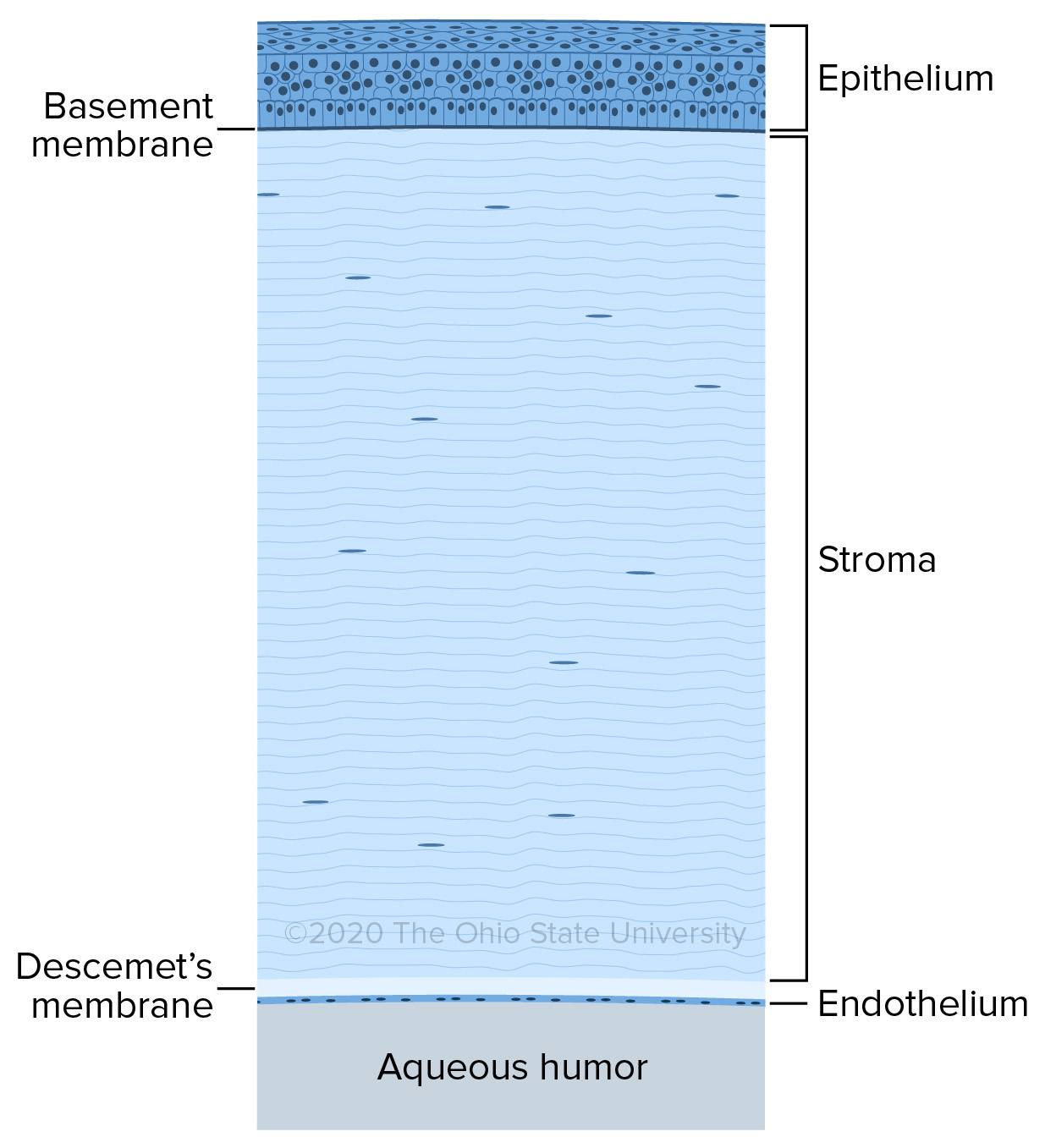

Sclera was divided into an episclera zone and a sclera proper zone.ĬONCLUSIONS: The outer layer of the episclera composed ofĬonnective fibers loosely attached to the sclera proper. Two distinct parts were separated by a distinct zone in addition, the RESULTS:TheĬornea of ostrich had both dermal and sclera components and the The sections were studied under a light microscope. Hematoxylin and eosin (H&E) and Masson's trichrome and PAS. Routine histological techniques were done and 6-μmthick Were kept in 10 % formalin solution for 7 days and then the eyes were All of them were inĪ good shape and healthy condition. Ostrich breeding center in Jupar, Kerman, Iran. METHODS: Ten mature ostriches were chosen from an Present study was to investigate the histology of the outer layer of That ostrich eyes would have distinct tissue structures and this has BACKGROUND: The Ostrich is an interesting subject concerningĪnimal evolution and morphology studies.


 0 kommentar(er)
